Breast Cancer Lymph Node Color Doppler Ultrasound Color Meanings

Normal Lymph Node On Ultrasound Lymph Nodes Typically Are Smooth Download Scientific Diagram
Color Doppler Sonography Characterizing Breast Lesions

Typical Us Appearance Of Lymph Nodes Typical Reactive Node Image A Download Scientific Diagram
Reactive Vs Malignant Lymph Nodes Ultrasound Features Radiology Reference Article Radiopaedia Org

Pdf Ultrasonography Of Superficial Lymph Nodes Benign Vs Malignant
Ultrasound Evaluation Of Regional Lymph Nodes As An Extension Of The Breast Ultrasound Exam Youtube
Thus for some time aln dissection alnd was the standard of care in these patients however screening programs and increased awareness on the disease tend to identify more breast cancer patients at an earlier stage in.
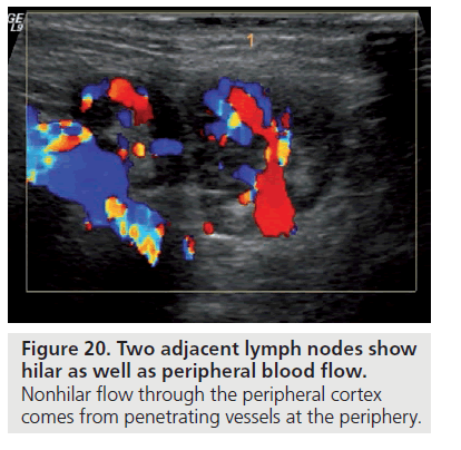
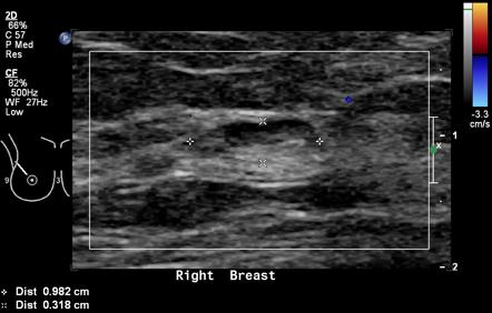
Breast cancer lymph node color doppler ultrasound color meanings.
Breast Cancer Topic Lymph Nodes On Ultrasound
Intramammary Lymph Nodes Radiology Reference Article Radiopaedia Org

Lymph Node Matting Multiple Malignant Nodes Are Fused In A Single Ill Download Scientific Diagram

Nodal Echogenic Hilum A Benign Node With Echogenic Central Hilum B Download Scientific Diagram
Https Journals Sagepub Com Doi Pdf 10 1177 8756479305278268

A 75 Year Old Woman With Primary Breast Lymphoma Ultrasound Image Of Download Scientific Diagram

Benign Node Oval Shape Preserved Central Hilum Download Scientific Diagram

Axillary Lymph Nodes In Breast Cancer Patients Sonographic Evaluation

Diagnostic Efficacy Of Color Doppler Ultrasound In Evaluation Of Cervical Lymphadenopathy Abstract Europe Pmc

An Axillary Lymph Node Showing Indeterminate Morphology On Conventional Download Scientific Diagram

Ultrasound Scanning Of The Pelvis And Abdomen For Staging Of Gynecological Tumors A Review Fischerova 2011 Ultrasound In Obstetrics Amp Gynecology Wiley Online Library

Three Dimensional Sonography Of Axillary Lymph Nodes In Patients With Breast Cancer Koenigsberg 2016 Journal Of Ultrasound In Medicine Wiley Online Library

Pdf Mistakes In Ultrasound Diagnosis Of Superficial Lymph Nodes
Http Pdf Posterng Netkey At Download Index Php Module Get Pdf By Id Poster Id 112347

Pdf Diagnostic Approach For Evaluation Of Lymph Node Metastasis From Thyroid Cancer Using Ultrasound And Fine Needle Aspiration Biopsy
Ultrasound Features Of Extranodal Extension In The Metastatic Cervical Lymph Nodes Of Papillary Thyroid Cancer A Case Control Study Mu Cancer Biology Medicine

The Role Of Preoperative Ultrasound Evaluation Of Inguinal Lymph Nodes In Patients With Vulvar Malignancy Sciencedirect

Pin On Breast Imaging
Https Encrypted Tbn0 Gstatic Com Images Q Tbn 3aand9gcs7s1f45hgr7vullszgzhelfimsjzsoglyqns9f0efp104yj3pr Usqp Cau

Pdf Review Of Ultrasonography Of Malignant Neck Nodes Greyscale Doppler Contrast Enhancement And Elastography
Evaluation Of Neck Lymph Node Metastasis On Contrast Enhanced Ultrasound An Animal Study
Ultrasound Features Of Thyroid Parathyroid Neck Lymph Nodes Normal And Pathologic Pattern Oncohema Key
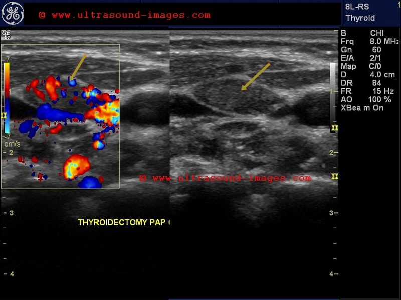
A Gallery Of High Resolution Ultrasound Color Doppler 3d Images Thyroid 2
Https Pubs Rsna Org Doi Pdf 10 1148 Rg 27si075502
Source : pinterest.com
