Breast Specimen Imaging Equipment

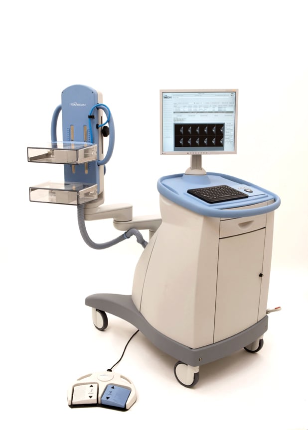
Trident System Specimen Radiography Hologic

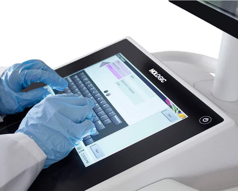
Brevera System Imaging And Verification Hologic

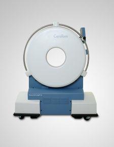
Mammotome Confirm System Core Specimen Radiography System Mammotome Neoprobe Sentimag
Emerging Trends In Breast Imaging Imaging Technology News

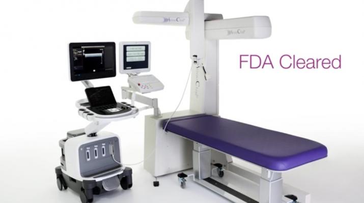
Mozart Specimen Tomosynthesis System Surgicalone

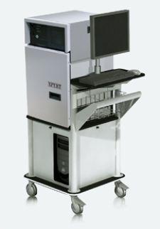
Specimen Radiography Products Kubtec Medical Imaging
Inspect integrated specimen tool 1 save time and costs.
Breast specimen imaging equipment.
Kubtec Imaging Systems

The Siemens 3t Skyra Mri Is Available In All Nine Of Our Zp Locations Radiology Imaging Home Appliances Home
Https Www Breverabiopsy Com Sites Brevera Files Brevera System Tech Specs Pdf

New Radiological Imaging Equipment Funded By Donation To San Foundation
Kubtec Has Specimen Radiography System Imaging Technology News
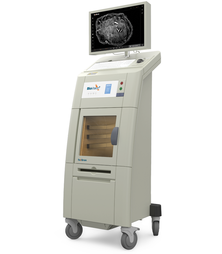
Biovision Radiation Cabinet Faxitron X Ray Sytem Products The Industrial Promoting Co Ltd

2003 Ge Voluson 730 Pro And 2005 Ge Voluson 730 Pro With No Transducer Available For Sale Direct With Medical Facility With Images Medical Equipment Pro Probe

Digital Mammography Equipment Price Cost Info

Ge Medical Imaging Equipment G E Walker Inc

T2 Mri Demonstrates A Very Thin Walled Cystic Lesion Which Follows Csf On All Sequences Features Are Characteristic Of An In Radiology Cysts Radiology Imaging

640px Crookes Tube Xray Experiment Jpg 640 405 X Ray Physics Vintage Medical
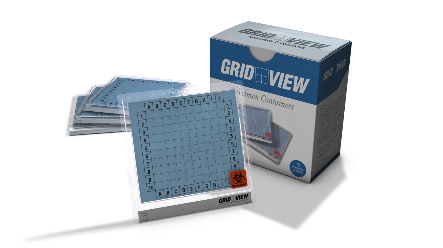
Grid View Specimen Imaging And Transport Container Cirs

Specimen Radiography System

Rtutec Detection Technology Used For Breast Cancer Specimen Imaging
Specimen Radiography For The Operating Room Kubtec Medical Imaging

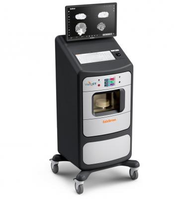
Fda Clears Faxitron Ct For 3d Specimen Radiography Axis Imaging News
Sonocine Releases 3 D Multiplanar Reconstruction Software To Improve Breast Cancer Detection Imaging Technology News

Specimen Radiography Beekley Medical
Rsna 1999 Diagnostic Imaging Equipment Manufacturers Unspool Digital Capabilities
Portable Scanner For X Ray Ct Applications Imaging Technology News

Alligator X Ray X Ray Art More At Fosterginger Pinterest Xray Art X Ray Animal Skulls

Qsum Channel Headrest Covers Fits A Range Of Headrests Shown On Website Mri Headrest Ultrasound

South Carolina Safe Surgery 2015 Circulating Nurse Hospitality Management Surgery
Hologic Acquires Digital Specimen Radiography Company Faxitron Bioptics Imaging Technology News
Source : pinterest.com
